What is The Test?
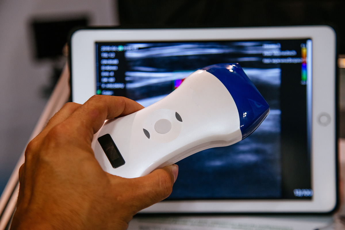
A pharmacological stress echocardiogram combines stress test with an echocardiogram. During a pharmacological stress echocardiogram, you will not exercise on a treadmill. In place of the treadmill exercise, a medication is used to increase your heart rate as if you were exercising. Ultrasound images of your heart are obtained at rest and again at intervals throughout out the exam. These resting and interval ultrasound images are compared to determine if coronary artery disease is present.
Patient Preparation
There is nothing to eat 4 hours before the test and no caffeine for 12 hours prior to the test. Medications may be taken unless told not to by your physician. Wear comfortable clothing.

What to Expect
Once in the testing room, we will ask you to sign a consent form to give us permission for the testing. An IV will be placed in your arm. You will be asked to remove your shirt and lie on the table on your left side. The initial resting echocardiogram is performed. The technologist will apply warm gel to your chest and use the transducer to create pictures of your heart on the ultrasound machine. The technologist will take several pictures and videos of your heart as measurements are obtained and your blood flow is checked. Four pre-stress pictures will then be obtained. Once the four pre-stress pictures are obtained, you will be connected to an EKG monitor and the pharmacological stress test will begin. A target heart rate will be calculated based on your age. This heart rate is necessary to meet for exam accuracy. Small doses of medication will be injected at intervals to increase your heart rate until the target is met. Ultrasound images of your heart are obtained at each interval. Once you have reached the target heart rate, the post-exercise pictures of your heart will be obtained and the exam is then ended. This exam will take approximately 60 minutes to complete. Our cardiologist will review the EKG stress test, resting echocardiogram and the pre-stress, interval and post-stress ultrasound images to determine the presence of coronary artery disease. The results of the exam will be discussed in your follow up visit.

